INTRODUCTION
There exists a dilemma within the clinical considerations involving the rehabilitation of the normal cervical lordosis. Should the doctor be utilizing ligamentous remodeling traction and postural neuromuscular re-education to reduce the forward head posture, or should the prudent clinician be concentrating his efforts on the correction of the normal cervical 63 degree arc of a circle primarily through the use of structural corrective extension traction.
While examining the pre and post radiographs of 30 consecutive patients treated with Ambulatory Postural Remodeling™ of the cervical spine, I have found some notable consistencies. These consistencies have helped me to develop certain objective guidelines for the decision to target my efforts toward the reduction of the patient’s aberrant forward head carriage or to concentrate on the restoration of his/her normal cervical lordosis.
In the course of this paper, I will describe Ambulatory Postural Remodeling™ of forward head posture, the consistencies in the reduction of forward head posture with different cervical curvatures and radiogaphic guidelines that will hopefully help you decide which type of cervical rehabilitation is best for your individual patient.
Ambulatory Postural Remodeling™
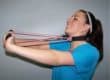
Ambulatory Postural Remodeling™ is a rehabilitative procedure I developed for the reduction of forward head posture. It involves having the patient wear a Cervical Remodeling Collar™ with the posterior cervical sling section in the more open settings while drawing the head back into retraction without inducing more than 15 to 20 degrees of head extension. The patient is encouraged to tuck his/her chin and try to hold the head level against the resistance of the forehead strap while he/she wears the collar (See Picture 1).
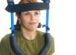
If the patient is physically able to walk on a treadmill at two to three miles per hour, he/she is instructed to do so while he/she wears the remodeling collar. This is done for eight to ten minutes and then the collar is taken off and replaced with a neoprene™ head weigh belt that can hold from one-half a pound to six pounds of lead weight in half-pound increments against the patient’s forehead (See Picture 2). It has been found that this will induce an involuntary neuromuscular head retraction for as long as the patient wears the head weight. The patient again walks on a treadmill at two to three miles per hour for eight to ten minutes while wearing the Posture Corrective Exercise Belt™. I am presently compiling 40 pre and post radiographs of consecutive patients treated with this ambulatory therapy and hope to publish the results in the near future.
An earlier clinical study of 30 consecutive patients who had 14-seven minute sessions of wearing the Cervical Remodeling Collar™ in a seated fashion while performing chin tuck exercises, revealed that such use of this device can reduce forward head posture by 8.43 millimeters in 77% of the subjects. This rate and ratio of postural correction are better than any other current method for the reduction of forward head posture.
Consistencies In The Reduction of Forward Head Posture
In the analysis of numerous pre and post cervical x-rays, first with forward head posture and then with reduced forward head translation, certain patterns can be identified. Patients can be grouped into true forward head posture (usually will be more than 30 millimeters), upper cervical forward head posture and lower cervical extension/ upper cervical forward head posture (both usually less than 30 millimeters). You will rarely also find patients with a retro head posture (See Figure A).
The points of reference for this analysis have been established by Harrison and Janik.1,2,3 Their peer-review published studies have established a normal global position for the cervical spine. Drs. Harrison and Janik’s studies indicate that the Cl plane line should ideally create a 28.7 degree angle to a horizontal plane line and that the intersection of posterior tangent lines drawn off C2-3 should intersect and create a 9.4 degree curve, C3-4/C4-5/C5-6 and C6-7 should intersect and create 8.2 degree curves. Anterior head translation should ideally be at zero. The cervical curve should be in a symmetrical circular lordosis about a 63-degree arc of a circle. Harrison CBP® Seminars produces and sells a plastic template that allows the doctor to easily produce the 63-degree arc line onto the patient’s lateral cervical x-ray and compare the patient’s current vertebral position to the ideal position.
My definition of true forward head posture is where any of the posterior bodies of the lower cervical vertebra (C4,5,6 or 7) are forward of the 63-degree arc line. Upper cervical forward head posture is where the posterior bodies of C4,5,6 and 7 are all on the 63-degree arc line or very close to it and Cl,2 and 3 are all forward of the arc line. The lower cervical extension/upper cervical forward head posture is where the posterior bodies of C4,5,6 and 7 are all behind the arc line and C1,2 and 3 are all forward of the arc line. Retro head posture is where the posterior bodies of the upper cervical segments are behind the 63-degree arc line.
I have found that by using my ambulatory method of cervical remodeling traction and neuromuscular re-education, I am able to reduce the forward head posture in all three of the forward head posture classifications I have just previously listed. I have also discovered that doing this procedure with patients who have the upper cervical forward head posture or the lower cervical extension/upper cervical forward head posture will usually have the adverse effect of drawing the lower cervical segments further into extension and away from their ideal position.
Objective Guidelines For Posture and Curve Correction
Because of my findings, I now recommend that the Cervical Remodeling Collar™ be used with the posterior cervical sling section in the more posterior (or open) positions only with patients who present with true forward head posture. Posture corrective head weighting should also only be performed by patients with true forward head posture. Once the posterior bodies of the patient’s lower cervical segments (C4,5,6,7) have reached the 63-degree arc line or are behind it, forward head posture correction procedures should stop and curve corrective extension traction should start. This will usually occur when the forward head translation is under 30 millimeters and almost always with cases of 15 millimeters or less.
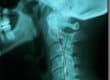
According to Doctor Deed Harrison in his CBP® Cervical Rehabilitation Seminar, the best form of extension traction for patients with a lower cervical extension/upper cervical flexion kyphotic curvature is two-directional extension compression traction. I have found that I can mimic this type of traction with the Cervical Remodeling Collar™ by first tightening the posterior cervical sling section all the way to the frame and placing it on the patient. I then use the forehead strap to draw the head and upper cervical spine into full extension over the posterior sling section (See Picture 3). If he/she is still able to walk while wearing the device in this matter, I have him/her do so on the treadmill for 10-12 minutes per session. Using the Remodeling Collar in this fashion, I am able to draw or maintain the posterior bodies of the lower cervical segments on the 63-degree arc line while the upper segments are traction back to the arc line. My post radiographic results using this new procedure have been very favorable and I highly recommend it for patients with only upper cervical forward head posture or lower cervical extension/upper cervical forward head posture.
REFERENCES
- Harrison DD, Janik TJ, Troyanovich SJ, Holland B. Comparisons of lordotic cervical spine curvatures to a theoretical ideal model of the static sagittal cervical spine. Spine 1996; 21:667-75.
- Harrison DD, Janik TJ. Clinical validation of an ideal normal static cervical spine model. In: Witten M, ed., Computational medicine, public health and biotechnology. Part 1. Austin, TX: World Scientific Publishing, 1995: 1036-46.
3. Janik TJ, Harrison DD. Prediction of 2-D static normal position of the cervical spine from mathematical modeling. In: Witten M, ed. Computational medicine, public health and biotechnology. Part 2. Austin, IX: World Scientific Publishing, 1995:1036-46.