INTRODUCTION
A condition of altered global joint position/posture commonly exists in the cervical spine. Approximately 66 percent of the population or more have the condition of anterior head translation either with or without symptoms.1 This structural aberrant posture is also known as head protrusion or forward head posture and has been linked to and associated with:
- tension, cerviogenic and migraine headaches.2 -9
- chronic neck and upper back pain with muscular overload/strain.10,11,12
- craniomandibular dysfunction, abnormal tooth wear patterns and craniofacial growth asymmetries and abnormalities.13
- craniocervical angle/motion and Obstructive Sleep Apnoea.14,15
- bone, disc, ligament and myofasial degeneration/pathology.16-23
- partly reversed curvature, especially in women.24
- increased tension of the spinal cord with structural dependent biomechanical deformations of the central nervous system.25,26,27 The increased tension seems to be a primary mechanism leading to neurological dysfunction. This is supported by several investigations concerning spinal column/spinal cord distraction and the relationship between axial tension in the spinal cord to neurological function.28-31 Because of the adverse effect forward head posture has on the nervous system, it has been suggested as a component in the etiology of thoracic outlet syndrome and dorsal scapular nerve entrapment.32,33
A recent study by Columbia University of 485 computer users, musicians, and others with work-related upper-extremity pain and other symptoms has found postural misalignment one of their significant findings with protracted shoulders in 78 percent of the patients and head forward position in 71 percent of the subjects. They concluded that despite initial presentation distally, work-related upper-extremity disorders are a diffuse neuromuscular illness with significant proximal upper-body findings that affect distal function.34
Because of the relationship between these numerous adverse conditions and forward head protrusion with cervical hypolordosis and the clinical need for a treatment that effectively
provides a reduction of the severity or a correction of these conditions, this paper has been written to demonstrate the clinical efficiency of a new therapy.
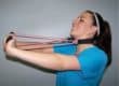
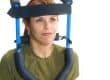
This ambulatory therapy is provided on a treadmill while the patient first wears a traction device that draws and holds their head into a position of posterior translation (head retraction) over an adjustable posterior cervical support, and then next wears an adjustable head weight (See Figure 1, A and B).
I have termed this therapy “Ambulatory Postural Remodeling.” The intent of this therapy is to reduce forward head posture while simultaneously encouraging the normal cervical lordosis and to re-balance the cervicothoracic neuro-musculature. This paper discusses its pre/post radiographic results in 25 consecutive cases.
METHODS
Subjects
For this clinical study, 25 consecutive cases that had all presented with non-acute cervical complaints and/or upper thoracic symptoms and/or head pain were selected from the records of a Fountain Valley, California chiropractic clinic.
Inclusion criteria for the treatment group required that the patients had completed a prescribed program of care consisting of treatment interventions two to three times per week for five to eight weeks. Additional inclusion criteria required that pre treatment and post treatment lateral cervical radiographs had to have all seven cervical vertebrae clearly visualized on the radiographs and the post radiographs could not be taken any sooner than 48 hours after the patient’s last treatment.
The number of 16 to 20 minute treadmill treatments varied from 12 to16 with an average of 14.36 traction/exercise sessions.
There were 13 males and 12 females in the treatment group with an average age of 45.8 years and a range of 17 to 68 years old. All subjects were from the Orange County, CA area.
Treatment Protocol
Chiropractic Biophysics® teaches a method of cervical analysis in which the patient’s posture is visually inspected and then correlated with the patient’s radiographic findings. The resultant spinal manipulation can be performed in a manual, diversified style with the patient in a prone position. The manipulation is delivered in a mirror-image direction to the abnormal posture found (ie. rotation, lateral flexion and translations). The patients received this type of manipulation on each treatment, usually after their ambulatory therapy.
The method of cervical traction used in this study is illustrated in Figure1A and is termed “negative Z-axis translation traction.” This was delivered via the use of a Cervical Remodeling Collar™ with the patient ambulatory at approximately 2-3 miles per hour on a standard, motorized treadmill. This wearable spinal orthosis allows for the application of an adjustable negative z-axis translation traction force to the patient’s skull while simultaneously providing an adjustable positive z-axis translation traction force to the patient’s upper, mid or lower cervical spine. The collar’s purpose is to reduce forward head posture while encouraging the return of a normal cervical lordosis.
In accordance with the Cervical Remodeling Collar instructions, the patient’s skull was first drawn into full retraction. Then the posterior cervical sling was tightened forward into the mid-cervical region. The sling was tightened to either patient tolerance or until the patient’s skull exhibited mild extension. The ambulatory traction usually started on the second treatment after no notable adverse physical reaction was noted from the patient’s first treatment of spinal manipulation. Five minutes of this therapy was performed on the first application and then the time was increased to eight to ten minutes over the next few sessions
Once the patients were experiencing only mild pain or no pain of their skull, cervical or upper thoracic regions; they were encouraged to slightly tuck their chin and endeavor to maintain their head level against the forehead strap. This active chin tuck exercise while in the traction collar is designed to increase the occipital/atlas separation and strengthen the anterior inferior cervical musculature. At this time, ambulatory head weighting was also initiated (Figure 1B).
The basic principle behind head weighting goes back to placing large books atop young ladies’ heads to help improve their posture. In 1975, Cailliet wrote of having the patient wear a two-to-ten-pound weight upon the head for varying periods during the day (10 to 30 minutes) to improve proper erect posture.35 The head weighting began at two pounds for five minutes. The length of this therapy was first gradually increased in one minute increments to eight to ten minutes and then the weight was gradually increased in one half pound increments to three to seven pounds
The patients were also given instruction in the performance of McKenzie head retraction exercises for daily home use. It is not known how well the patients complied with their home exercise program.
Measurement Procedures
Lateral cervical radiographs were obtained using a Chiro-Tech 250-125 x-ray machine, rare earth screens, and an automatic exposure meter to minimize patient exposure. All of the subjects were standing for the lateral cervical view and were instructed to flex and extend their head with their eyes closed and then assume a neutral, comfortable position. The patient’s head was only adjusted to endeavor to keep the hard palate level and eliminate axial rotation on the pre and post x-rays.
A x-ray technician with two years of CBP® experience served as radiographic examiner and made a series of geometric measurements on both the pre and post radiographs.
These measurements included determination of the angular relationships of adjacent vertebrae from C2 through C7 (ie., C2-3, C3-4, C4-5, C5-6, C6-7).
These adjacent segmental measurements shall be referred to as relative rotation angles. The relationship of the plane of C1 to a horizontal reference line was also measured and shall be referred to as the atlas plane angle.
The illustration in Figure 2 depicts measurement of the relative rotation angles from C2 through C7. In actually drawing these lines, the examiner drew posterior vertebral body lines through the posterior-superior body corner and the posterior-inferior body corner of each vertebra of C2 through C7.
Instead of measuring a relative rotation angle between C1 and C2, a line representing the atlas plane is compared to horizontal and drawn through the inferior anterior arch and inferior posterior tubercle of C1. In addition to these six angles, anterior head translation was measured in millimeters. This measurement is an anterior-posterior displacement of C2 to C7, as shown in Figure 2.
Displacement of the posterior-superior body margin of C2 is measured relative to a vertical axis line (VAL) drawn through the posterior-inferior body margin of C7. The method of line drawing analysis used in this study has been described in a prior study by Jackson et al.36 The reliability of this method of radiographic measurement was established in this study.
In regard to the cervical spine, a normal global position has been documented in the peer- review literature. Dr. Harrison and Janik’s studies37,38,39 indicate that the C1 plane line should ideally create a 28.7 degree angle to a horizontal plane line and that the intersection of posterior tangent lines drawn off C2-3 should intersect and create a 9.4 degree curve, C3-4/C4- 5/C5-6 and C6-7 should intersect and create 8.2 degree curves. Anterior head translation should ideally be at zero in a symmetrical circular lordosis about a gravity line.
In Table 1, my pre/post x-ray data is analyzed as to whether the resultant vertebral position moved closer or further away from the ideal value, the average amount of change and the occurrence of relative rotation angle increase or decrease per spinal level.
RESULTS
As the patient data illustrates in Tables 1, I found this manipulation/ traction/exercise program to reduce forward head posture in 96 percent of the subjects. The amount of reduction in these patients ranged from two millimeters to 18 millimeters with an average of 10.32 millimeters of reduced forward head posture. This last number includes one patient whose forward head posture remained unchanged after the treatment.
The C1 angle improved in 76 percent of the patients. The amount of C1 angle increase in these patients ranged from one degree to10 degrees with an average of 3.16 degrees. This last number includes six patients whose C1 angle reduced further on the post radiograph.
The relative rotation angles moved closer to the ideal value 40 percent of the time, stayed the same 22.67 percent of the time and either decreased below the ideal value or increased over the ideal value 37.33 percent of the time. The largest percentage of relative rotation angular increase was observed at the C1, C4/C5, C5/C6 and C6/C7 levels. The most frequent occurrence of relative rotation angular decrease was seen at the C2/C3 and C3/C4 spinal levels. The C1 spinal level was the site of most change, much more often increasing than decreasing. The C2/C3 and C3/C4 spinal levels were the only relative rotation angles to have the average post treatment result be a decrease of curvature. All other levels resulted in an increase of curvature as the post treatment effect with an overall average of 5.16 degrees of increased cervical curvature.
CONCLUSION
Being able to reduce forward head translation in 96 percent of your patients, by an average of ten millimeters, while increasing their cervical lordosis by an average of five degrees after just 14 treatments is superior to any comparable rehabilitative treatment in the literature. This therapy warrants further research into its short and long term effectiveness in the treatment of the many posture related physical disorders listed at the beginning of this paper.
REFERENCES
- Griegel-Morris P, Larson K, Mueller-Klaus K, Oatis CA. Incidence of common postural abnormalities in the cervical, shoulder, and thoracic regions and their association with pain in two age groups of healthy subjects. Phys Ther 1992; 72:425-31.
- Campbell DG, Parsons CM. Referred head pain and its concomitants. J Nerv Ment Dis. 1944; 99;544-51.
- Jensen R, Rasmussen BK. Muscular disorders in tension-type headache. Cephalalgia 1996; 16:97-103.
- Watson DH, Trott PH. Cervical headache: an investigation of natural head posture and upper cervical flexor muscle performance. Cephalalgia 1993; 13:272-284.
- Kidd RF, Nelson R. Musculoskeletal dysfunction of the neck in migraine and tension headache. Headache 1993; 33:566-9.
- Mercer S, Marcus DA, Nash J. Cervical musculoskeletal disorders in migraine and tension-type headache. Paper presented at the 68th annual meeting of the American Physical Therapy Association. 1993; Cincinnati, Ohio.
- Marcus DA, Scarf L, Mercer S, Turk DC. Musculoskeletal abnormalities in chronic headache: a controlled comparison of headache diagnostic groups. Headache 1999;39:21-7.
- Vernon H, Steinman I, Hagen C. Cervicogenic dysfunction in muscle contraction headache and migraine: a descriptive study. J Manipulative Physiol. The 1992; 16:428-31.
- Nagasawa A, Sakakibara T, Takahashi A. Roentgenographic findings of the cervical spine in tension-type headache. Headache 1993; 33:90-5.
- Griegel-Morris P, Larson K, Mueller-Klaus K, Oatis CA. Incidence of common postural abnormalities in the cervical, shoulder, and thoracic regions and their association with pain in two age groups of healthy subjects. Phys Ther 1992; 72:425-31.
- Cailliet R. Neck and arm pain. Philiadelphia: F. A. Davis; 1981.
- Haughie LJ, Fiebert IM, Roach KE. Relationship of forward head posture and cervical backward bending to neck pain. J Manipulative Physiol Ther 1995; 3:91-97.
- Gonzalez H, Manns A. Forward head posture: its structural and functional influence on the stomatognathic system, a conceptual study. J Craniomandib Pract 1996; 14:71-80.
- Ozbek MM, Miyamoto K, Lowe AA, Fleetham JA. Natural head posture, upper airway morphology and obstructive sleep apnoea severity in adults. Eur J Orthod 1998; 20:133-43.
- Tangugsorn V, Skatvedt O, Krogstad O, Lyberg T. Obstructive sleep apnea: a cephalometric study, part I. cervico-craniofacial skeletal morphology. Eur J Orthod 1995; 17:45-56.
- Stillwelll D. Structural deformities of vertebrae: bone adapatation and modeling in experimental scoliosis and kyphosis. J Bone Joint Surg Am 1962; 44:611-33.
- Adams MA, McMillan DW, Green TP, Dolan P. Sustained loading generates stress concentrations in lumbar intervertebral discs. Spine 1996; 21:434-8.
- Fukuyama S, Nakamura T, Takagi K. The effect of mechanical stress on hypertrophy of the lumbar ligamentum flavum. J Spinal Disord 1995; 8:126-30.
- Zagra A, Lamartina C, Pace A. Posterior spinal fusion in scoliosis: computer-assisted tomography and biomechanics of the fusion mass. Spine 1998; 13:155-9.
- Jones M, Pais J, Omiya B. Bony overgrowths and abnormal calcifications about the spine. Radiol Clin North Am 1988; 26:1213-34.
- Stokes IA, Spence H, Aronsson DD, Kilmer N. Mechanical modulation of vertebral body growth: implications for scoliosis progression. Spine 1996; 21:1162-7.
- Horst, Brinckmann P. Measurement of the distribution of axial stress on the end-plate of the vertebral body. Spine 1981; 6:217-31.
- Matsunaga S, Sakou T, Taketome E, Nakanisi K. Effects of strain distribution in the intervertebral discs on the progression of ossification of the posterior longitudinal ligaments. Spine 1996; 21:184-9.
- Visscher CM, de Boer W, Naeije M. The relationship between posture and curvature of the cervical spine. J Manipulative Physiol Ther 1998; 21:388-91.
- Breig A. Adverse mechanical tension in the central nervous system. Analysis of cause and effect. Relief by functional neurosurgery. New York: John Wiley and Sons; 1978.
- Smith C. Changes in length and position of the segments of the spinal cord with changes in posture in the monkey. Radiology 1956; 66:259-65.
- Ruch WJ. Atlas of common subluxations of the human spine and pelvis. Boca Raton (FL): CRC Press; 1997.
- Fujita Y, Yamamoto H. An experimental study on spinal cord traction effect. Spine 1989; 14:698-705.
- Naito M, Owen J, Bridwell K, Sugioka Y. Effects of distraction of the physiologic integrity of the spinal cord, spinal cord blood flow, and clinical status. Spine 1992; 17:1154-8.
- Yamada S, Zinke D, Sanders D. Pathophysiology of tethered cord syndrome. J Neurosurg 1981; 54:494-503.
- Yamada S, Iacono R, Andrade T, Mandybur G, Yamada B. Pathophysiology of tethered cord syndrome. Neurosurg Clin N Am 1995; 6:311-23.
- Smith KF. The thoracic outlet syndrome: a protocol of treatment. J Orthop Sports Phys Ther 1979; 1:89-99.
- Darnell MW. A proposed chronology of events for forward head posture. J Craniomandibular Prac 1983; 1:49-54.
- Pascarelli EF, Hsu YP. Understanding work-related upper extremity disorders: clinical findings in 485 computer users, musicians, and others. J Occup Rehabil 2001 Mar; 11 (1):1-21.
- Cailliet R. Scoliosis, diagnosis and management. Philadelphia: F.A. Davis Company; 1975.
- Jackson BL, Harrison DD, Robertson GA, Barker WF. Chiropractic Biophysics lateral cervical film analysis reliability. J Manipulative Physiol Ther 1993; 16:384-91.
- Harrison DD, Janik TJ, Troyanovich SJ, Holland B. Comparisons of lordotic cervical spine curvatures to a theoretical ideal model of the static sagittal cervical spine. Spine 1996; 21:667-75.
- Harrison DD, Janik TJ. Clinical validation of an ideal normal static cervical spine model. In: Witten M, ed., Computational medicine, public health and biotechnology. Part 1. Austin, TX: World Scientific Publishing, 1995: 1036-46.
- Janik TJ, Harrison DD. Prediction of 2-D static normal position of the cervical spine from mathematical modeling. In: Witten M, ed. Computational medicine, public health and biotechnology. Part 2. Austin, TX: World Scientific Publishing, 1995:1036-46.